Pupillary Hippus as a Component of Post-Attenuation Neurological Signs in a Dog
Monday September 16, 2024
Last week, our neurologist, Max Foreman, showcased this case study, Pupillary Hippus as a Component of Post-Attenuation Neurological Signs in a Dog, in a poster presentation at the ECVN conference in Portugal. Max, an EBVS-recognised specialist in Veterinary Neurology and Neurosurgery, qualified from the University of Cambridge in 2015. His career includes experience at Wanstead Veterinary Hospital and Dick White Referrals, where he completed his residency and earned the European Diploma in Neurology and Neurosurgery. Max has also contributed extensively to research, presenting at both national and international conferences.
Introduction
Post-attenuation neurological signs (PANS) are a recognised complication following surgical attenuation of congenital extrahepatic portosystemic shunts (EHPSS) in dogs. These neurological signs can manifest within days after surgery and are distinct from hepatic encephalopathy. While PANS commonly involve symptoms like seizures, behavioural changes, or ataxia, the occurrence of pupillary hippus as a component of PANS is particularly rare. This article presents a unique case of pupillary hippus following shunt surgery in a dog, highlighting its significance and clinical management.
What is Pupillary Hippus?
Pupillary hippus, also referred to as pupillary athetosis or pupillary nystagmus, is an exaggerated, cyclic motion of the pupils, where they constrict and dilate rhythmically without any changes in external stimuli such as light, emotional state, or focus. Although well-documented in human medicine, where it has been linked to conditions like non-convulsive status epilepticus and vestibular migraines, its occurrence in veterinary patients is much less understood.
Case Presentation
A four-year-old neutered male Pug was referred for evaluation after presenting with polyuria, polydipsia, sporadic vomiting, and hyporexia. Bile acid stimulation testing revealed elevated concentrations of both pre-prandial and post-prandial bile acids, prompting further investigation. A computed tomography angiogram confirmed the presence of a left gastro-azygos EHPSS.
Following two weeks of medical management, the dog underwent surgical placement of a 5mm ameroid constrictor around the shunt. The initial recovery was uneventful, but 48 hours post-surgery, the dog developed severe neurological symptoms, including tonic-clonic seizures and cortical blindness. Examination revealed rhythmic, oscillatory dilation and constriction of both pupils—consistent with a diagnosis of pupillary hippus.
Management and Treatment
To control the seizures, diazepam and levetiracetam were administered intravenously. Despite this, two additional tonic-clonic seizures occurred, prompting the introduction of phenobarbitone and a dexmedetomidine continuous rate infusion to manage the dog’s dysphoria and vocalisation. Supportive care included lactulose, omeprazole, and metoclopramide. No further seizures were observed after the addition of phenobarbitone, and the pupillary hippus resolved within 24 hours. Over the following eight weeks, the dog made a full neurological recovery.
Discussion
Pupillary hippus is a rare but noteworthy component of PANS. In human medicine, it is often associated with non-convulsive status epilepticus, a possible consideration in this case. While electroencephalography (EEG) was not performed, it is plausible that the hippus could have represented seizure activity. However, as the neurological signs, including the hippus, resolved independently over time, it remains possible that the hippus was a self-limiting and separate neurological sign unrelated to seizure activity.
This case demonstrates the importance of recognising less common neurological signs like pupillary hippus in dogs with PANS. While the exact mechanism behind pupillary hippus remains unclear, its presence can serve as a valuable clinical indicator of underlying neurological disturbances following EHPSS surgery. Early and appropriate management is crucial to improving outcomes in such cases.
Conclusion
The occurrence of pupillary hippus as part of post-attenuation neurological signs in a dog undergoing surgical attenuation of an EHPSS is a rare and intriguing presentation. This case underscores the need for awareness of the full spectrum of neurological signs that may accompany PANS. Further research into the mechanisms behind pupillary hippus and its role in post-surgical recovery could enhance veterinary understanding and improve the management of dogs with PANS.
DOWNLOAD POSTER HERE
Download pdfAlso in the news
New study identifies variation in BOAS risk across brachycephalic breeds
Cuneiformectomy for Advanced Laryngeal Collapse – What the Latest Evidence Means for Your BOAS Patients

Statement on the CMA’s Investigation into the Veterinary Sector
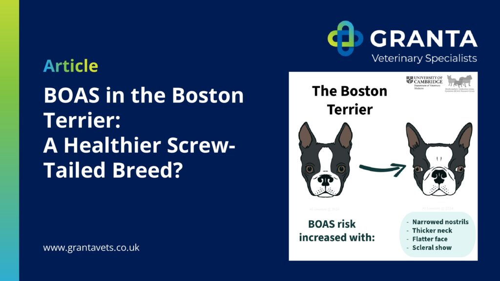
BOAS in the Boston Terrier: A Healthier Screw-Tailed Breed?
Please note that our turnaround time for outpatient medication requests has increased to 5 working days. This applies to both written prescriptions and the dispensing of physical medications.
We appreciate your patience and understanding while we process requests as efficiently and safely as possible. If you have any urgent queries, please contact the team directly.
Thank you for your ongoing support.