Neural network analysis of pharyngeal sounds can detect obstructive upper respiratory disease in brachycephalic dogs.
Monday September 30, 2024
Groundbreaking Neural Network Detects BOAS in Brachycephalic Dogs
BOAS (Brachycephalic Obstructive Airway Syndrome) is a serious respiratory condition affecting breeds like French Bulldogs and Pugs. A new study co-authored by Jane Ladlow introduces a neural network model that detects BOAS with remarkable accuracy. This technology analyses stertor (a low-frequency snoring sound), a key symptom of BOAS, using recordings from electronic stethoscopes, achieving an 85% accuracy rate. This automated approach provides a more objective diagnosis compared to human-based methods.
BOAS in Brachycephalic Breeds
BOAS is widespread among brachycephalic (flat-faced) breeds, affecting 60% of Pugs and 50% of French Bulldogs. It leads to various health issues, including poor exercise tolerance, sleep disorders, and reduced lifespan. Diagnosis traditionally relies on subjective assessment of respiratory noise, often resulting in variable outcomes. The neural network, developed by McDonald, Ladlow, and colleagues, introduces automation and consistency. It successfully detects and grades stertor by analysing 665 recordings from 341 dogs, achieving 71% sensitivity and 86% specificity, helping provide earlier and more consistent diagnoses.
Key Figures from the Study
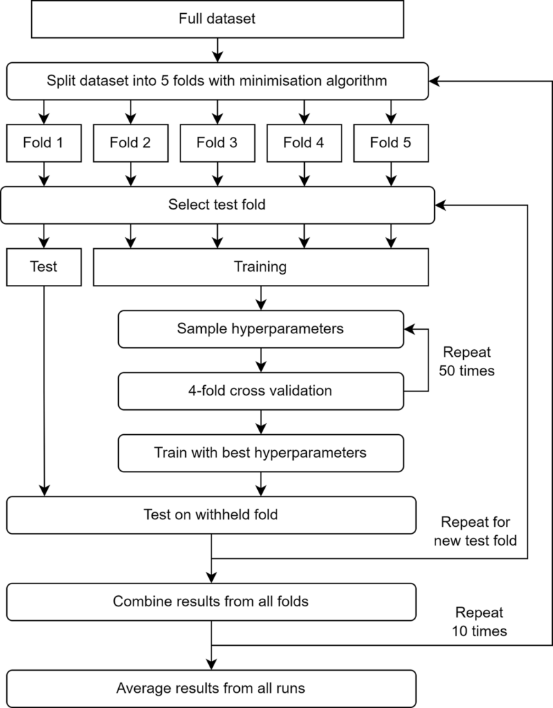
- Figure 1 details the experimental setup, explaining how the recordings were processed through the neural network using a nested cross-validation approach. This ensures the reliability and generalisability of the model.
- Figure 2 provides a spectrogram of a healthy French Bulldog’s respiratory sound with no audible stertor or stridor, showcasing normal breathing patterns.
- Figure 3 shows the spectrogram of a Pug with moderate BOAS, clearly illustrating multiple stertor sounds, visible as strong low-frequency tonal sounds.
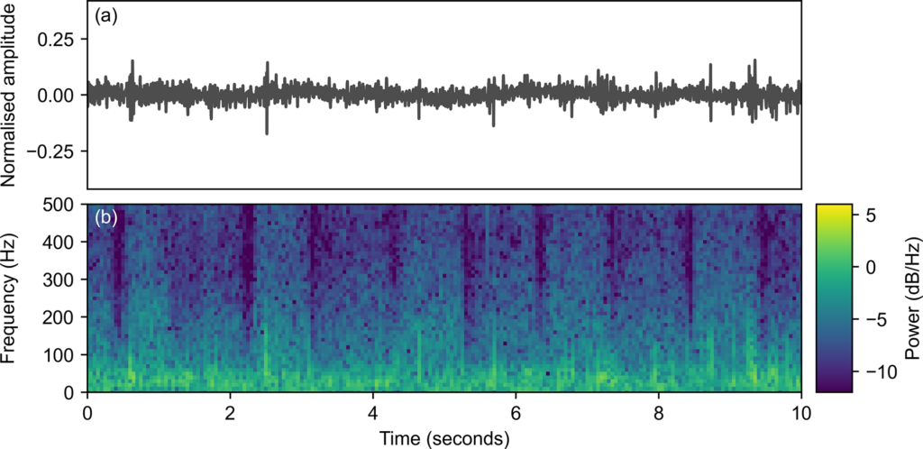

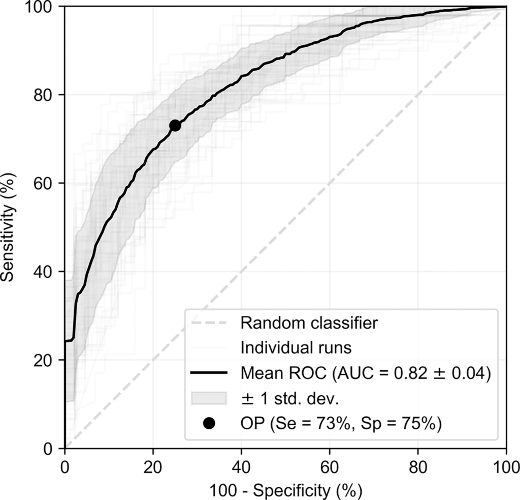
These visual insights provide a look at how the neural network distinguishes between normal and BOAS-affected dogs, making the study both scientifically robust and visually informative.
BOAS Airway Clinic at Granta Veterinary Specialists
The BOAS Airway Clinic specialises in the diagnosis and treatment of BOAS, offering solutions ranging from lifestyle modifications to tailored surgical interventions. Early detection of BOAS can prevent complications, helping brachycephalic dogs lead healthier, more comfortable lives.
Future of BOAS Detection
This neural network marks a major breakthrough in veterinary diagnostics, with the potential to extend beyond clinics and into homes. As the technology advances, it could be implemented in a smartphone app linked to electronic stethoscopes, allowing pet owners to monitor their dogs’ respiratory health at home. This would enable earlier detection and timely intervention.
Moreover, future enhancements of the model could focus on detecting stridor, another indicator of airway obstruction, further improving the accuracy of BOAS diagnosis.
Learn More and Download the Study
For more information about BOAS diagnosis and treatment, visit our BOAS Airway Clinic page.
To explore the full details of this groundbreaking research, download the paper from PLOS One here.
Also in the news
New study identifies variation in BOAS risk across brachycephalic breeds

Cuneiformectomy for Advanced Laryngeal Collapse – What the Latest Evidence Means for Your BOAS Patients

Statement on the CMA’s Investigation into the Veterinary Sector